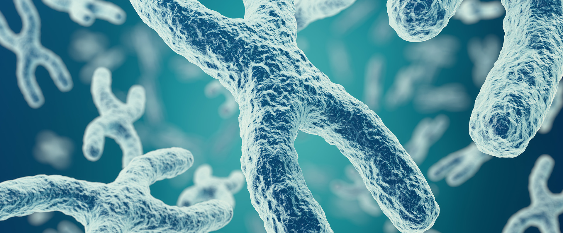
Aneuploidy refers to the presence or absence of one or more additional chromosomes. As each chromosome is composed of hundreds of genes, the addition or deletion of a single chromosome disrupts the equilibrium of the cell and, in the majority of cases, is incompatible with life. Down syndrome and Turner syndrome are common examples of aneuploidy disorders. Therefore, screening tests such as ultrasound, NIPT, and microarray during pregnancy are recommended as they give information about the baby’s risk of developing a chromosomal disorder.
How does chromosomal aneuploidy occur?
Nondisjunction (Errors in Meiosis)
The most common cause of aneuploidy is nondisjunction, which occurs when the chromosomes do not separate correctly during the cell division process. Nondisjunction usually occurs in meiosis I, where a chromosome pair does not divide in the egg or in the sperm cell, resulting in two cells having an extra chromosome and two cells having a missing chromosome.
Nondisjunction also occurs in meiosis II, where the sister chromatid (one half of a duplicated chromosome) will not divide, resulting in one cell having an extra chromosome, one cell with a missing chromosome, and two cells with the right number of chromosomes.
Trisomies and Monosomies
|
A trisomy occurs when a sperm or an egg cell combines with one of these extra chromosome cells. A trisomy results in three extra chromosomes instead of the usual two. A monosomy occurs when a sperm or egg cell combines with a missing chromosome cell, and the resultant cell (zygote) will have only one chromosome.
Robertsonian translocation
Other than nondisjunction, Robertsonian translocation accounts for a small proportion of aneuploidies. Each chromosome is split into sections (shorter arm and longer arm) based on the position of a narrowing (constriction) called a centromere.
Robertsonian translocation occurs in acrocentric chromosome, where the centromere is present at the very end of the chromosome. If two acrocentric chromosomes break at their centromeres, then the two long arms can fuse to form a single chromosome with both long arms, or the two short arms can fuse to make a smaller chromosome with two short arms.
Since the long arms carry the most genetic material, the cell with the long arm will essentially have an additional chromosome, which, when combined with a normal reproductive cell, will lead to trisomy. Whereas cells with short arms almost lack a chromosome and can lead to monosomy when combined with normal reproductive cells.
Types of chromosomal aneuploidies
Autosomal aneuploidies
The majority of aneuploidy affecting autosomal chromosomes is life-threatening and typically results in spontaneous abortion during the first trimester. There are three viable autosomal aneuploidies:
Down syndrome: This is caused due to the presence of three copies of chromosome 21 and affects approximately 1 in every 700 babies. Down’s syndrome is characterized by a distinctive facial structure, developmental delay, intellectual disability, thyroid disease, or cardiac disease.
Edwards syndrome: This is caused due to the presence of three copies of chromosome 13 and affects approximately 1 in every 8,000 babies. The majority of fetuses with Edwards syndrome die prior to birth, while those who survive the pregnancy typically die within weeks to months of birth as a result of complications. Infants with Edwards syndrome are characterized by dysmorphic features, including small eyes, a small head, a cleft lip, etc. Additionally, they may suffer from intellectual disabilities, developmental delays, and heart, kidney, and gastrointestinal disorders.
Patau syndrome: This is caused due to the presence of three copies of chromosome 18 and affects approximately 1 in 15,000 babies. The majority of fetuses with Patau syndrome die before or after birth as a result of complications. Patau syndrome is characterized by severe intellectual disability, deformities of the skull and nose, polydactyly (extra fingers), etc.
Sex chromosome aneuploidies
Sex chromosome aneuploidies are generally not life-threatening due to the lesser genetic contribution of X and Y chromosomes to overall growth and development in an individual. Two common aneuploidies affecting the sex chromosomes include Klinefelter syndrome and Turner syndrome.
Klinefelter syndrome: This is caused when a fetus with the XY genotype inherits one or more extra X-chromosomes. The most common characteristics of Klinefelter syndrome are male sterility, small external genitals, long legs and a short torso, scarce facial and body hair, and gynecomastia (the growth of breast tissue). This condition commonly affects males.
Turner syndrome: This is caused when one of the X-chromosomes is missing completely or partially in a fetus. Turner syndrome is characterized by non-functional fibrous ovaries, short stature, neck webbing, and congenital heart and kidney defects. This condition affects only females.
How is aneuploidy diagnosed?
Microarray testing
- Microarray, also known as chromosomal microarray (CMA) is a widely used method for detecting fetal chromosomal aneuploidy by examining fetal blood or tissue, amniocytes, and chorionic villi. This test compares a fetal specimen to a standard sample to detect extra or missing chromosome fragments that are undetectable under a microscope. Microarray testing utilizes a microchip platform, enabling the simultaneous analysis of multiple pieces of DNA.
Microarray test result interpretation
- Normal: No missing or extra pieces of chromosomes were found.
- Likely pathogenic: Presence of a missing or extra piece of chromosome that may result in intellectual or developmental problems.
- Variant of unknown significance (VUS): Presence of a missing or extra piece of chromosome, but it is unclear whether this will lead to intellectual or developmental problems. VUS is a common occurrence.
- Regions of Homozygosity (ROH): One or many parts of the chromosome are genetically identical.
- Noninvasive prenatal testing (NIPT) NIPT is a noninvasive test that uses chromosomal microarray (CMA) to look for fetal chromosomal imbalances. This test detects the presence of the baby’s genetic material in the mother’s bloodstream. It is a screening test done during pregnancy to determine if the fetus has a risk of developing a genetic disorder like Down syndrome, Edwards syndrome, Patau syndrome, etc. NIPT is not a diagnostic test; therefore, the results of this test should be confirmed with subsequent cytogenetic follow-up tests.
Interpreting NIPT results:
- A positive NIPT test result indicates that the fetus may be at an increased risk of developing chromosome abnormalities.
- A negative NIPT test result indicates that the fetus has a reduced likelihood of developing a chromosomal disorder.
Errors in chromosome segregation lead to aneuploidy. Aneuploidy is more likely to occur during pregnancy in women of advanced maternal age and can be diagnosed using chromosomal microarray (CMA) testing. Although extensive research has been conducted over the years, there are currently no treatments available for aneuploidies. Consequently, the management of aneuploidy depends on individual requirements and associated complications.


